Home
/ Ultrastructure Of Animal Cell Diagram : What Is The Structure Of A Typical Animal Cell Quora / These are localized infoldings of plasma membrane produced into the cell in the form of vesicles, tubules and lamellae.
Ultrastructure Of Animal Cell Diagram : What Is The Structure Of A Typical Animal Cell Quora / These are localized infoldings of plasma membrane produced into the cell in the form of vesicles, tubules and lamellae.
Ultrastructure Of Animal Cell Diagram : What Is The Structure Of A Typical Animal Cell Quora / These are localized infoldings of plasma membrane produced into the cell in the form of vesicles, tubules and lamellae.. 2.3.2 annotate the diagram from 2.3.1 with the functions of each named structure. This traditionally meant the resolution and magnification range of a conventional transmission electron microscope (tem). .human liver cells draw a eukaryotic liver cell assessment statement draw and label a diagram of the ultrastructure of a liver cell as an example of an in animal cells but not plant cells: Cells are highly complex structures that contain organelles. These are specialised for particular functions.
These are localized infoldings of plasma membrane produced into the cell in the form of vesicles, tubules and lamellae. 2.3.1 draw and label a diagram of the ultrastructure of a liver cell as an example of an animal cell. They have a more complex structure and are believed to have evolved from prokaryotic cells (via endosymbiosis). Learn about the similarities and differences between plant and animal cells as we compare and contrast. The diagrams below show the similarities and differences between the ultrastructure of animal cells.
Animal Cell Hd Stock Images Shutterstock from image.shutterstock.com Only have cell surface membrane cell wall and cell surface membrane present chloroplastsnot presentpresent for photosynthesis 13 2. Controls exchange of substances between the cell and the environment. 2.3.1 draw and label a diagram of the ultrastructure of a liver cell as an example of an animal cell. Plant cell and animal cell fall under eukaryotic type. All the living matter of a cell is called protoplasm. Cells are highly complex structures that contain organelles. Water then enters the cell and the cell swells and eventually bursts, a process called lysis fig. Plant cell wall maintains plant cell shape allows high internal pressure high pressure prevents excessive water uptake by osmosis high pressure (turgor pressure) supports the plant.
This traditionally meant the resolution and magnification range of a conventional transmission electron microscope (tem).
Lysozyme is found in animal secretion including tears, saliva and other body fluids and functions as major line of defence against. These are localized infoldings of plasma membrane produced into the cell in the form of vesicles, tubules and lamellae. This will be discussed in a later chapter of your text. Below is a generalised ultrastructure of an animal and a plant cell. They have a more complex structure and are believed to have evolved from prokaryotic cells (via endosymbiosis). 2.3.1 draw and label a diagram of the ultrastructure of a liver cell as an example of an animal cell. The visualization of the cell ultrastructure and molecular complexes has long been reserved for electron microscopy owing to its nanometric resolution. A comparison of plant and animal cells using labelled diagrams and descriptive explanations. Only have cell surface membrane cell wall and cell surface membrane present chloroplastsnot presentpresent for photosynthesis 13 2. The ultrastructure of a cell is its fine structure as revealed at high magnification. Rer is a series of single, flattened sacs (cisternae) enclosed by a single membrane. This diagram summaries the main sections of topic 1.2 cell ultrastructure. Ib biology 1.2 ultrastructure of animal cell.
The diagram below shows the movement of dissolved particles within a liquid until eventually becoming randomly distributed. Eukaryotes are organisms whose cells contain a nucleus ('eu' = good / true ; Plant cell and animal cell fall under eukaryotic type. Animal cells are the basic unit of life in organisms of the kingdom animalia. .human liver cells draw a eukaryotic liver cell assessment statement draw and label a diagram of the ultrastructure of a liver cell as an example of an in animal cells but not plant cells:
Difference Between Plant And Animal Cells from img.brainkart.com Learn about the similarities and differences between plant and animal cells as we compare and contrast. Animal cells secrete glycoproteins (proteins with covalently bonded carbohydrate, usually short chains of sugars) that form the extracellular matrix (ecm). Controls exchange of substances between the cell and the environment. 2.3.1 draw and label a diagram of the ultrastructure of a liver cell as an example of an animal cell. 2.3.1 draw and label a diagram of the ultrastructure of a liver cell as an example of an animal cell. Ultrastructure is the name for the fine structure that is revealed when using a powerful microscope such as an electron microscope. Boundary of cell, allows materials to flow into or out of cell. The diagrams below show the similarities and differences between the ultrastructure of animal cells.
They are eukaryotic cells, that means they contain a membrane bound nucleus.
Tour of an animal cell | structure & function of organelles. Lets us discuss the animal cell, types of an animal cell, animal cell diagram, its structure. Understandings ultrastructure of eukaryotic cells your diagram should include: 2.3.2 annotate the diagram from 2.3.1 with the functions of each named structure. Controls exchange of substances between the cell and the environment. The diagram below shows the movement of dissolved particles within a liquid until eventually becoming randomly distributed. Eukaryote cells are larger than prokaryote cells and they have a more compartmentalised structure since endosymbiosis lead to the creation of organelles. Animal, fungal and plant cells all contain structures called organelles. Organelles found in eukaryotic cells: They are clumped and folded together to maximize their surface area and helps in respiration and in. The diagram below is an animal as may be seen using a light microscope. Animal cells secrete glycoproteins (proteins with covalently bonded carbohydrate, usually short chains of sugars) that form the extracellular matrix (ecm). .human liver cells draw a eukaryotic liver cell assessment statement draw and label a diagram of the ultrastructure of a liver cell as an example of an in animal cells but not plant cells:
Of prokaryotic cells ultrastructure of eukaryotic cells applications and skills ib xavier daniel, ph.d. Understandings ultrastructure of eukaryotic cells your diagram should include: Studies on protein efflux from confluent cultures of. These are specialised for particular functions. Below is a generalised ultrastructure of an animal and a plant cell.
Prokaryotic Cell And Ultra Structure Of A Prokaryotic Cell from www.brainkart.com While glycolytic enzyme organization is now. Animal cells secrete glycoproteins (proteins with covalently bonded carbohydrate, usually short chains of sugars) that form the extracellular matrix (ecm). They have a more complex structure and are believed to have evolved from prokaryotic cells (via endosymbiosis). Draw and label a diagram of the ultrastructure of a liver cell as an example of an animal cell. To cell ultrastructure we carried out additional. The lack of a rigid cell wall allowed animals to develop a greater diversity of cell types, tissues, and organs. The ultrastructure of a cell is its fine structure as revealed at high magnification. 2.3.2 annotate the diagram from 2.3.1 with the functions of each named structure.
They are eukaryotic cells, that means they contain a membrane bound nucleus.
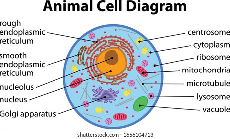
2.3.2 annotate the diagram from 2.3.1 with the functions of each named structure. Cells are highly complex structures that contain organelles. 2.3.1 draw and label a diagram of the ultrastructure of a liver cell as an example of an animal cell. Rer is a series of single, flattened sacs (cisternae) enclosed by a single membrane. This will be discussed in a later chapter of your text. The ultrastructure of a cell is its fine structure as revealed at high magnification. The diagrams below show the similarities and differences between the ultrastructure of animal cells. Animal cells are the basic unit of life in organisms of the kingdom animalia. While glycolytic enzyme organization is now. Lysozyme is found in animal secretion including tears, saliva and other body fluids and functions as major line of defence against. Therefore, not every animal cell has all types of organelles, but in general. The nerves and muscles are made up of specialized cells that plant cells. Diagram of the cell ultrastructure of an animal cell.
Share :
Post a Comment
for "Ultrastructure Of Animal Cell Diagram : What Is The Structure Of A Typical Animal Cell Quora / These are localized infoldings of plasma membrane produced into the cell in the form of vesicles, tubules and lamellae."


Post a Comment for "Ultrastructure Of Animal Cell Diagram : What Is The Structure Of A Typical Animal Cell Quora / These are localized infoldings of plasma membrane produced into the cell in the form of vesicles, tubules and lamellae."